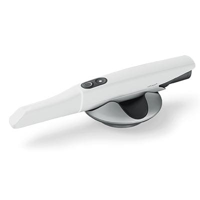
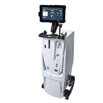
PIC System Implant Oral Scanner
The PIC (Precise Implants Capture) System is a cutting-edge oral scanner designed specifically for taking digital impressions of multiple dental implants.
Key benefits:
- Precise implant position and angle
- Digital alternative to traditional methods
- Useful for full arch, All-On-4 dental implants, where accuracy is critical for a successful outcome.
Navident Implant Placement Guidance System
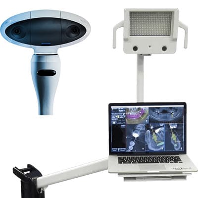
The Navident Implant Placement Guidance System is dentistry's newest dynamic navigation system. It provides real-time, 3D guidance during implant placement to enhance the precision and accuracy of dental implant surgeries.
Key benefits:
- Improves the accuracy of implant placement
- Helps avoid critical anatomical structures, reducing the risk of complications
- Ensures predictable and successful treatment outcomes
- Enables minimally invasive procedures
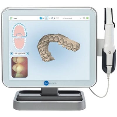
3Shape Trios 5 Dental Scanner
3Shape Trios 5 is a state-of-the-art tool that Captures highly accurate digital impressions of a patient's mouth and empowers dentists to provide efficient, accurate, and comfortable care.
Key benefits:
- Generats detailed and accurate 3D models of the teeth and gums
- Captures realistic color and texture
- Integrates with other dental software and equipment
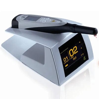
Millennium PerioLase MVP-7 Dental Laser
The Millennium PerioLase MVP-7 is a cutting-edge dental laser specifically designed to treat periodontal (gum) disease. This laser is used in the LANAP® (Laser-Assisted New Attachment Procedure) protocol, a minimally invasive alternative to traditional gum surgery.
Key benefits:
- Targets the bacteria associated with gum disease
- Promotes faster healing and regeneration of new tissue.
- Helps regenerate new cementum, periodontal ligament, and alveolar bone.
- Reduces bleeding and swelling associated with the procedure
Kavo Diagnodent Laser Caries Detection Unit
The KaVo DIAGNOdent is a laser fluorescence device used in dentistry to detect dental caries (tooth decay) at its earliest stages before dentists can see it or even on X-rays.
Key benefits:
- Detects caries at an earlier stage vs traditional methods
- Provides a complete and accurate assessment of decay
- Non-invasive and painless procedure.
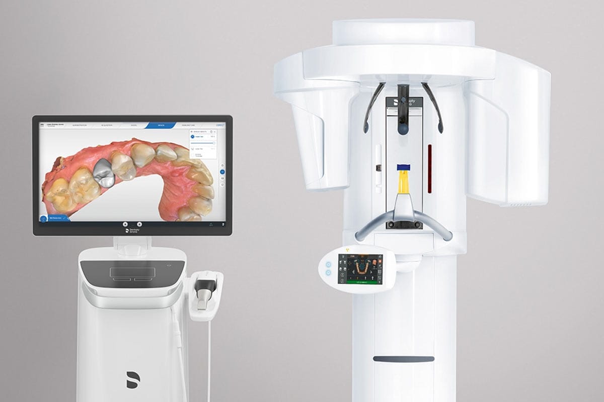
Dentsply Sirona Orthophos CBCT
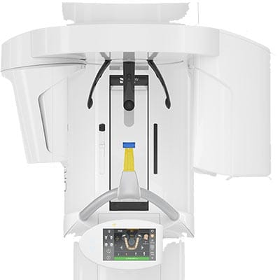
The Dentsply Sirona Orthophos CBCT is a sophisticated dental imaging system that uses Cone Beam Computed Tomography (CBCT) technology to create detailed 3D X-ray images of a patient's teeth, jaws, and surrounding facial structures. CBCT is commonly used in Implant Dentistry, Cosmetic Dentistry, Restorative Dentistry, Oral Surgery, Endodontics, and Orthodontics.
Key benefits:
- Comprehensive 3D Imaging
- High-resolution detail images
- Minimizes radiation exposure
Penguin RFA - Implant Stability Monitor
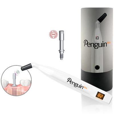
The Penguin RFA (Resonance Frequency Analysis) is used in implant dentistry to measure the stability of dental implants. It helps dentists determine when an implant is ready to support a restoration (like a crown, bridge, or denture).
Key benefits:
- Improve the predictability and success of implant restoration
- Assess implant stability and detects early signs of implant instability
- Determine the optimal time to attach dental crown, bridge, or denture
3D Seiler Dental Surgical Microscope
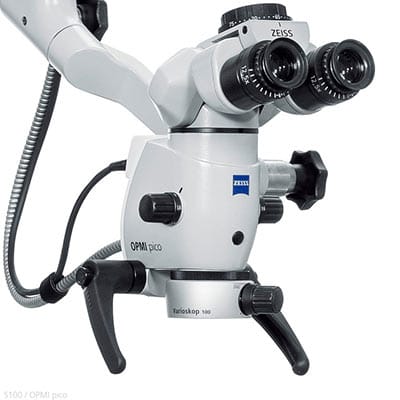
A 3D Seiler dental surgical microscope is a high-powered microscope specifically designed for dentistry. It provides magnified, three-dimensional, enhanced visualization for a wide range of dental procedures.
Key benefits:
- Improves visualization of root canal
- Enhances precision in tooth preparation for fillings, crowns, and other restorations.
- Improves visualization during Periodontal (Gum) Disease treatment
- Enhances precision during implant placement
MicronMapper S.I.N. 360 Photogrammetry
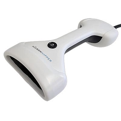
The MicronMapper S.I.N. 360 helps achieve accurate measurements of dental implant positions, especially in complex full-arch implant cases, such as All-On-4, Full Mouth Dental Implants, and Denture Implants.
Key benefits:
- Provides exceptional precision in measuring implant positions
- Integrates with various dental labs for the implant fabrication
- Quick and efficient scanning process
Schick USBCam4 Intraoral Camera
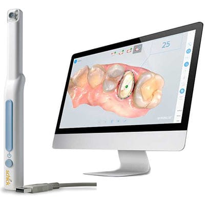
The Schick USBCam4 an intraoral camera captures images and videos inside a patient's mouth. It provides a view of the teeth, gums, and other oral structures.
Key benefits:
- Provides clear, high-resolution images that allow dentists to see fine details
- Lightweight and easy to handle, making it comfortable for both the dentist and the patient
- Integrates with major dental imaging and practice management software
Dentrix Electronic Claims Submission
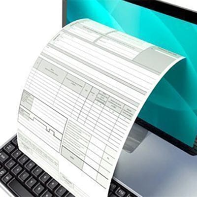
Dentrix software streamlines the process of submitting dental insurance claims, making it faster, more efficient, and less prone to errors compared to traditional paper claims.
Key benefits:
- Quicker reimbursement from insurance companies
- Saves time by automating the claims process
- better claim status visibility
For more information about the technologies we use in our dentistry center, call us at (516) 352-1000. To meet any of our dentists, visit us in Franklin Square, in the heart of the Town of Hempstead.
Clock Tower Dental
110 New Hyde Park Rd
Franklin Square, New York 11010
516-352-1000